Hemostasis Practical Manual⁚ A Comprehensive Guide
This manual serves as a practical, succinct guide for clinicians seeking quick answers to everyday questions. It offers a comprehensive overview of hemostasis and thrombosis, guiding readers through diagnosis, management, and prevention of hemorrhagic and thrombotic diseases. It includes essential practical management for all professionals working in the field of hemostasis and thrombosis.
Introduction to Hemostasis
Hemostasis, the process by which the body stops bleeding, is a complex and tightly regulated physiological mechanism involving a coordinated interplay of vascular, platelet, and coagulation factors. It is essential for maintaining the integrity of the circulatory system and preventing life-threatening blood loss following injury. The process is initiated by vascular injury, triggering a cascade of events that culminate in the formation of a stable hemostatic plug, effectively sealing the damaged blood vessel. This intricate process involves vasoconstriction, platelet adhesion and aggregation, and the activation of the coagulation cascade, leading to the formation of a fibrin clot.
Hemostatic disorders can arise from deficiencies in any of these components, leading to either excessive bleeding or inappropriate clotting. Excessive bleeding, or hemorrhage, can result from deficiencies in platelet function, coagulation factors, or vascular integrity. Conversely, inappropriate clotting, or thrombosis, can occur when the hemostatic system becomes overactive, leading to the formation of blood clots within blood vessels, potentially obstructing blood flow and causing tissue damage. Understanding the intricacies of hemostasis is crucial for diagnosing and managing a wide range of clinical conditions, from minor cuts and bruises to life-threatening thromboembolic events.
This manual aims to provide a comprehensive overview of hemostasis, including its physiological mechanisms, laboratory tests, and clinical implications. It will delve into the intricacies of the coagulation cascade, exploring the role of various coagulation factors and their interactions. Additionally, it will discuss common hemostatic disorders, their clinical presentations, and the latest diagnostic and therapeutic approaches. By providing a practical guide to the diagnosis and management of hemostatic disorders, this manual serves as an invaluable resource for clinicians and laboratory personnel working in the field of hemostasis and thrombosis.

The Coagulation Cascade
The coagulation cascade is a complex series of enzymatic reactions that ultimately leads to the formation of a stable fibrin clot, essential for hemostasis. It involves a precise interplay of various coagulation factors, primarily synthesized in the liver, which act as proenzymes that are sequentially activated in a tightly regulated manner. The cascade is traditionally divided into three pathways⁚ the intrinsic, extrinsic, and common pathways, each with its unique initiation and activation mechanisms. The intrinsic pathway is triggered by contact activation, involving factors XII, XI, IX, and VIII, while the extrinsic pathway is initiated by tissue factor, a protein released from damaged cells, activating factor VII.
Both the intrinsic and extrinsic pathways converge at the common pathway, where factor X is activated, leading to the generation of thrombin. Thrombin, a key enzyme in the coagulation cascade, converts fibrinogen into fibrin monomers, which then polymerize to form fibrin polymers. The fibrin polymers, along with aggregated platelets, form the stable hemostatic plug. The coagulation cascade is a highly regulated process, involving a delicate balance between procoagulant and anticoagulant factors, ensuring efficient hemostasis without excessive clotting. Several mechanisms control the coagulation cascade, including the presence of natural anticoagulants such as antithrombin, protein C, and protein S, as well as the rapid clearance of activated clotting factors from circulation.
Understanding the intricacies of the coagulation cascade is essential for diagnosing and managing hemostatic disorders. This knowledge allows clinicians to interpret laboratory tests, identify deficiencies in specific coagulation factors, and tailor treatment strategies to address underlying hemostatic abnormalities. The manual will delve deeper into the specific mechanisms of each pathway, highlighting the role of individual coagulation factors, their interactions, and the regulatory mechanisms that govern this vital process.
Laboratory Tests of Hemostasis

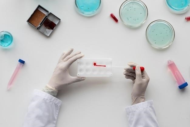
Laboratory tests play a crucial role in the evaluation of hemostasis, providing valuable insights into the functionality of the coagulation cascade, platelet function, and overall hemostatic balance. These tests are essential for diagnosing bleeding disorders, monitoring anticoagulation therapy, and identifying potential risks for thrombosis. The most commonly performed tests include⁚
Complete Blood Count (CBC)⁚ This test measures the number of red blood cells, white blood cells, and platelets in the blood, providing a basic assessment of blood cell count, including platelets, which are essential for primary hemostasis.
Prothrombin Time (PT)⁚ This test assesses the extrinsic pathway of coagulation by measuring the time it takes for plasma to clot after the addition of tissue thromboplastin, a reagent that activates factor VII.
Activated Partial Thromboplastin Time (aPTT)⁚ This test evaluates the intrinsic pathway of coagulation by measuring the time it takes for plasma to clot after the addition of a reagent that activates factor XII.
International Normalized Ratio (INR)⁚ This standardized measure of PT, calculated using a specific formula, is used to monitor anticoagulation therapy, particularly for patients on warfarin, a vitamin K antagonist.
Fibrinogen Assay⁚ This test measures the level of fibrinogen in the blood, a crucial protein involved in clot formation.
Platelet Aggregation Studies⁚ These tests evaluate the ability of platelets to aggregate and form a primary hemostatic plug in response to various stimuli, such as collagen, adenosine diphosphate (ADP), and epinephrine.
This manual will provide a comprehensive guide to the principles, interpretation, and clinical significance of these laboratory tests, empowering clinicians to effectively utilize these tools for diagnosing, monitoring, and managing hemostatic disorders.
Hemostatic Disorders
Hemostatic disorders encompass a spectrum of conditions that disrupt the delicate balance between bleeding and clotting, leading to either excessive bleeding or inappropriate thrombosis. These disorders can arise from inherited genetic defects or acquired conditions, impacting the intricate interplay of blood vessels, platelets, and coagulation factors.
Bleeding disorders, characterized by an increased tendency to bleed, can result from deficiencies in coagulation factors, platelet dysfunction, or abnormalities in vascular integrity. Common examples include⁚
Hemophilia A and B⁚ These inherited disorders involve deficiencies in factor VIII (hemophilia A) or factor IX (hemophilia B), leading to prolonged bleeding episodes, particularly after trauma or surgery.
Von Willebrand Disease⁚ This most common inherited bleeding disorder affects von Willebrand factor, a protein essential for platelet adhesion and coagulation factor VIII stability.
Thrombocytopenia⁚ This condition, characterized by a low platelet count, can result from various causes, including autoimmune disorders, medications, and infections, leading to increased bleeding risk.
Conversely, thrombotic disorders, characterized by an increased propensity for blood clot formation, can arise from genetic predisposition, acquired conditions, or a combination of factors.
Deep Vein Thrombosis (DVT)⁚ This condition involves blood clots forming in deep veins, typically in the legs, potentially leading to pulmonary embolism.
Stroke⁚ This serious condition occurs when a blood clot blocks an artery in the brain, interrupting blood flow and damaging brain tissue.
Understanding the underlying mechanisms and specific characteristics of these disorders is crucial for accurate diagnosis, effective treatment, and appropriate management of bleeding and thrombotic complications.
Hemophilia
Hemophilia, an inherited bleeding disorder, arises from a deficiency in specific clotting factors, primarily factor VIII (hemophilia A) or factor IX (hemophilia B). These proteins play crucial roles in the coagulation cascade, a complex series of reactions that ultimately leads to the formation of a stable fibrin clot, halting bleeding.
Individuals with hemophilia experience prolonged bleeding episodes, often following minor injuries, surgeries, or even spontaneous bleeding into joints. The severity of hemophilia varies depending on the level of clotting factor deficiency. Severe hemophilia, with low factor levels, often leads to spontaneous bleeding, while milder forms may only manifest with prolonged bleeding after trauma.
The diagnosis of hemophilia typically involves a thorough medical history, physical examination, and laboratory tests. Blood tests are used to measure the levels of clotting factors, confirming the diagnosis and determining the severity of the disorder.
Treatment for hemophilia focuses on preventing and controlling bleeding episodes. Prophylactic treatment, involving regular infusions of the deficient clotting factor, can significantly reduce the frequency and severity of bleeds. On-demand treatment, where clotting factor is administered only during bleeding episodes, is also available but may be less effective in preventing complications.
Advances in gene therapy and other novel therapies hold promise for long-term management and potentially even cures for hemophilia. These developments offer hope for individuals with hemophilia, aiming to improve their quality of life and reduce the burden of this lifelong condition.
Von Willebrand Disease
Von Willebrand disease (VWD) is a common inherited bleeding disorder affecting both genders. Unlike hemophilia, which primarily affects males, VWD can occur in both sexes. The disease stems from a deficiency or dysfunction of von Willebrand factor (VWF), a protein crucial for platelet adhesion and coagulation. VWF acts as a bridge, connecting platelets to damaged blood vessels, initiating the clotting process.
Individuals with VWD experience a range of bleeding symptoms, from mild to severe, depending on the severity of the VWF deficiency. Common manifestations include easy bruising, prolonged bleeding from minor cuts, nosebleeds, heavy menstrual bleeding in women, and gastrointestinal bleeding. In severe cases, spontaneous bleeding may occur.
Diagnosis of VWD involves a thorough medical history, physical examination, and specialized blood tests. Laboratory tests evaluate VWF levels, its function, and platelet aggregation. These tests help determine the type and severity of VWD, guiding treatment strategies.
Treatment for VWD primarily focuses on controlling bleeding episodes. Desmopressin, a synthetic hormone, can temporarily increase VWF levels in some individuals. VWF concentrates, derived from human plasma, can be administered to replace the deficient VWF. For individuals with severe VWD, prophylactic treatment with VWF concentrates may be necessary to prevent spontaneous bleeding.
Managing VWD involves a multidisciplinary approach, with close collaboration between hematologists, primary care physicians, and other healthcare professionals. Early diagnosis and appropriate treatment can significantly improve the quality of life for individuals with VWD, minimizing bleeding complications and promoting overall well-being.
Thrombosis
Thrombosis refers to the formation of a blood clot (thrombus) inside a blood vessel, obstructing blood flow. This potentially life-threatening condition can occur in both arteries and veins, leading to various complications depending on the location and size of the clot. Arterial thrombosis can cause heart attack, stroke, or peripheral artery disease, while venous thrombosis can lead to deep vein thrombosis (DVT) and pulmonary embolism (PE).
Several factors can contribute to thrombosis, including inherited genetic predispositions, acquired conditions like cancer or autoimmune diseases, and lifestyle factors such as smoking, obesity, and prolonged immobility. Certain medications, like oral contraceptives or hormone replacement therapy, can also increase the risk of thrombosis.
Diagnosis of thrombosis often involves a combination of clinical assessment, imaging studies, and blood tests. Physical examination may reveal signs of swelling, pain, or redness in the affected area. Imaging tests, such as ultrasound, CT scan, or MRI, can visualize the clot and assess its location and extent. Blood tests, such as D-dimer and coagulation studies, can help confirm the presence of a clot and assess the risk of future thrombosis.
Treatment for thrombosis depends on the location and severity of the clot. Anticoagulant medications, such as heparin or warfarin, are commonly used to prevent further clot formation and allow existing clots to dissolve. In some cases, thrombolytic therapy, which involves administering medications that dissolve existing clots, may be necessary. In certain situations, surgical procedures, such as thrombectomy or angioplasty, may be required to remove the clot or restore blood flow.
Preventing thrombosis involves addressing modifiable risk factors, such as maintaining a healthy weight, quitting smoking, engaging in regular physical activity, and managing underlying medical conditions. For individuals at high risk, prophylactic anticoagulation therapy may be recommended.
Anticoagulants
Anticoagulants are medications that prevent the formation of blood clots or break down existing clots. These medications are crucial in the management of thrombotic disorders, such as deep vein thrombosis (DVT), pulmonary embolism (PE), stroke, and heart attack. Anticoagulants work by inhibiting various steps in the coagulation cascade, thereby reducing the ability of the blood to clot.
There are several different types of anticoagulants, each with its unique mechanism of action and clinical applications. Heparin, a naturally occurring substance found in mast cells, is a commonly used anticoagulant. It works by enhancing the activity of antithrombin, a naturally occurring protein that inhibits coagulation factors. Heparin is typically administered intravenously or subcutaneously and is often used in acute settings, such as the treatment of DVT or PE.
Warfarin, a synthetic vitamin K antagonist, is another widely prescribed anticoagulant. It inhibits the production of vitamin K-dependent clotting factors, such as factor II, VII, IX, and X. Warfarin is administered orally and is often used for long-term prevention of thrombotic events. It requires regular blood monitoring to ensure therapeutic levels and prevent bleeding complications.
Direct oral anticoagulants (DOACs), such as dabigatran, rivaroxaban, apixaban, and edoxaban, have emerged as newer alternatives to traditional anticoagulants. They directly inhibit specific coagulation factors, such as thrombin or factor Xa, without requiring routine blood monitoring. DOACs are generally well-tolerated and offer convenient oral administration, making them suitable for long-term anticoagulation therapy.
The choice of anticoagulant depends on the specific clinical situation, including the type of thrombotic event, patient characteristics, and potential drug interactions. Careful patient selection, monitoring, and management are crucial to optimize anticoagulation therapy and minimize the risk of bleeding complications.
Need to pass your Alaska driver’s exam? We’ve got you covered! Download the official Alaska DMV manual & practice tests – free & easy to use. Get licensed with confidence!
Lost your 2024 Lincoln Nautilus manual? Find everything you need – from maintenance to features – right here! Easy access & instant answers. **Lincoln Nautilus** made simple.


